Testicular Ultrasound Training
Hands-On Scrotal & Testicular Ultrasound Imaging Training
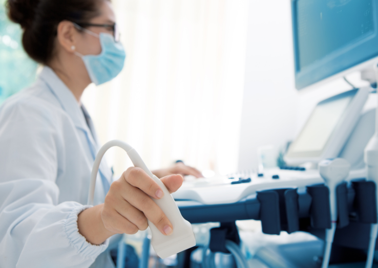
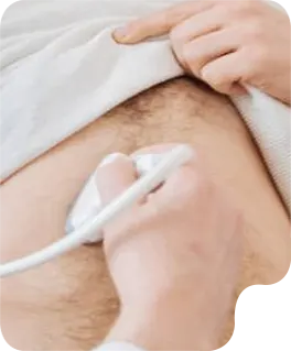
A focused, hands-on training program designed to build expertise in scrotal and testicular ultrasound imaging. Learn proper scanning techniques, anatomy identification, and pathology recognition with real clinical examples. Ideal for sonographers and healthcare professionals seeking accuracy, confidence, and diagnostic excellence.
The left testicle is the other primary male reproductive gland, situated within the scrotal sac, where it produces sperm and testosterone. Ultrasound imaging of the left testicle is essential for evaluating its overall health, detection conditions like swelling, infection, or the presence of lesions.
The left epididymis head is located at the top of the epididymitis Associated with he left testicle, serving s the initial collection point for sperm. Ultrasound examination of the left epididymis head is performed to detect issues such as epididymitis, hydroceles, or other palpable masses.
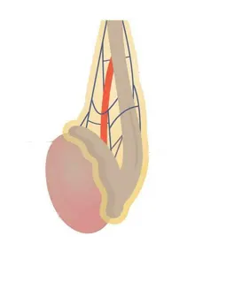
Doppler ultrasound of the left testicle assesses the blood Flow dynamics within the left testicular parenchyma and surrounding vessels. It is invaluable for detecting conditions such as left testicular torsion, where blood flow is acutely absent or diminished, or for identifying varicose veins(varicocele) impacting the left pumping form plexus.
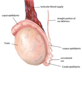
The left epididymis body extends along the back surface of the left testicle, serving as a conduit and storage area for maturing sperm. Ultrasound of the left epididymis body helps identify pathologies like inflammation, benign cystic formations, or abnormalities causing scrotal pain.
The right epididymis body is the central, elongated portion of the epididymis running along the posterior aspect of the right testicle. Ultrasound allows for detailed assessment of this segment, looking for signs of epididymitis, epididymis cysts, or other inflammatory changes
Doppler ultrasound of the right testicular retries and vines. this technique is critical for diagnosing conditions like testicular torsion, which involves compromised blood supply, or inflammatory processes indicated by increase blood flow.
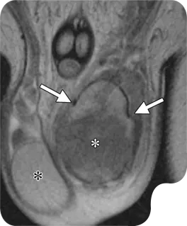
The right testicle is one of the to male productive glands located within the scoutmaster responsible for producing sperm a male hormones. Ultrasound provides detailed images to assess its size, internal texture, and any potential abnormalities such as cysts, masses, or signs of inflammation.
The right epididymis head is the uppermost part of the coiled The right epididymis head is the uppermost part of the coiled tube(epididymis) positioned on the superior aspect of the right testicle. Ultrasound helps visualize this structure to identify any fluid collection, inflammation (epididymitis), or small cysts.
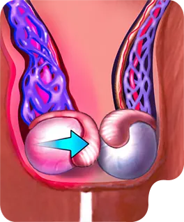
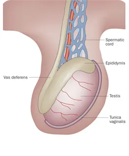
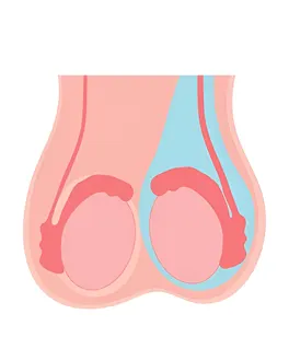
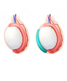
This ultrasound examination focuses on the scrotum, the sac that contains the testicles, epididymis, and the spermatic cord. It is used to evaluate for abuses of course pain, swelling, or masses, such as testicular torsion, epididymitis, o hydroceles. Doppler imaging can be added to assess blood flow within the testicles, which is crucial in cases of suspected torsion.
EXCELLENTTrustindex verifies that the original source of the review is Google. I recently had the pleasure of visiting Gentle Touch Sono Lab for an ultrasound, and I cannot express how impressed I was with the entire experience. I was able to meet Ms Penn. From the moment I entered her warm, welcoming office, I felt at ease. Ms. Penn's professionalism and expertise were immediately evident, putting me at comfort during what can often be a stressful time.She took the time to explain the procedure thoroughly and answered all my questions with patience and clarity. The ultrasound itself was conducted with the utmost care, and I appreciated her attention to detail throughout the process. Her state-of-the-art equipment ensured that I received excellent images, and she made sure to share the results with me in a way that was easy to understand.What truly stood out was Ms. Penn's compassionate nature. She took the time to connect with me on a personal level, making sure I felt supported and informed every step of the way. I highly recommend Ms. Penn to anyone in need of ultrasound services. Her combination of skill, empathy, and a calming atmosphere makes her the go-to professional in this field. Thank you, Ms. Penn, for a wonderful experience!Posted onTrustindex verifies that the original source of the review is Google. Gentle Touch Sono Lab helped me enhance my skills in vascular. I was struggling with lower extremity Arterial. After a few sessions, I feel much more confident .Posted onTrustindex verifies that the original source of the review is Google. WOW. I was speechless, it was such a beautiful space filled with so many training tools. I had the pleasure of meeting Ms. Penn, she was so sweet & knowledgeable & just so passionate about the craft !! I would definitely recommend any & everyone who might be interested in sonography to go check it out.Posted onTrustindex verifies that the original source of the review is Google. I had a great experience at Gentle Touch Sono Lab. The staff was friendly, professional, and made me feel comfortable throughout the entire visit. The sonographer explained everything clearly and took their time, which I really appreciated. The lab was clean, organized, and my appointment started on time. I would definitely recommend Gentle Touch Sono Lab to anyone looking for a comfortable and professional ultrasound experience.Posted onTrustindex verifies that the original source of the review is Google. I highly recommend Gentle Touch Lab! I originally came in not knowing where to start, and Felicia made sure I received the one-on-one support I needed to excel. I’ve gained a lot of experience from both Felicia and Louis. I’ll definitely be recommending them to all my radiology classmates.
Hands-on ultrasound training for students and professionals. Bridging the gap between learning and real-world practice.
Other Links
Contact Info
- +1 888-283-6907
- info@greenyellow-crane-487162.hostingersite.com
- 41B W Merrick Rd Suite 2, Valley Stream, NY 11580, United States